EMG/NCS Online Series: Volume I
Electronic Myoanatomic Atlas for Clinical Electromyography ( 2nd Edition )
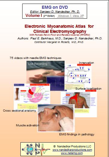
Proper identification and activation of muscles is critical for assessment of needle EMG recordings. Dr Barkhaus methodically explains muscle identification using the anatomic landmarks and palpation. Upon identifying the site of needle insertion, live EMG signals are seen as the subject activates and relaxes the muscle. This collection of 75+ videos covers the routinely tested muscles from upper limb, lower limb, neck, face and back (paraspinal). Dr Margret Roberts make an important contribution by discussing the pelvic floor muscles. Muscles used for chemo-denervation and some uncommon muscles are also included. The pictures show surface and cross sectional anatomy. A textbook in electronic format is also included for those who like printed copies.
Author(s) : Paul E. Barkhaus, M.D.; Sanjeev D. Nandedkar, Ph.D.
Contributor(s) : Margaret M. Roberts, M.D., Ph.D.
Compatibility : Windows PC, MAC PC, Mac Book, iPad, iPhone, Smart Phones, Tablets and other devices
Pricing Options Availability: In Stock Continue ShoppingDescription
- 70 Digital Video Clips of the commonly tested muscles with Needle EMG techniques
- 300+ still images with anatomical landmarks
- Female Pelvic Floor EMG studies
- Surface localization
- Cross sectional anatomy
- Anatomic data such as roots, innervation etc.
- Live recordings
- EMG findings in pathology
- Muscle activation
- 100+ page book, with practical input, ready to be printed
- Use it on a Multimedia Windows XP-SP2 PC
- Ideal teaching tool for academic centers
- Great reference material for practicing EMGers
- DVD media offers instantaneous access to the requested muscle data.
- No time consuming rewinding or fast forwarding of the video tapes!

Samples
EMG on DVD Series: Volume I
Electronic Myoanatomic Atlas for Clinical Electromyography
With Female Pelvic Floor and Related Muscles (PFARM)Authors Paul E. Barkhaus, M.D.; Sanjeev D. Nandedkar, Ph.D.
Contributors Margaret M. Roberts, M.D., Ph.D.
Video Samples
Frontalis
Infraspinatus